
 | ||
Romanowsky staining is a prototypical staining technique that was the forerunner of several distinct but similar methods, including Giemsa, Jenner, Wright, Field, and Leishman stains, which are used to differentiate cells in pathologic specimens. It was named after the Russian physician Dmitri Leonidovich Romanowsky (1861–1921), who invented it in 1891.
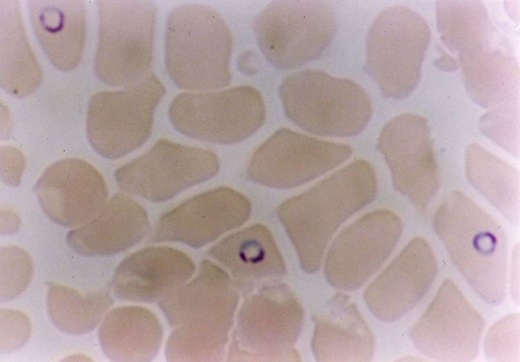
Microscopic examination of stained blood films
Paul Ehrlich had used mixtures of acidic and basic dyes for this purpose in 1879: e.g. fuchsine (acid dye) and methylene blue (basic dye). In 1891, Romanowsky developed techniques using a mixture of eosin Y and modified methylene blue that produced a surprising hue unattributable to either staining component: a beautiful, distinctive shade of purple. Requirement for the occurrence of the Romanowsky-Giemsa effect are:
- A cationic dye: The best dye is azure B and, though azure A gives the nuclear purple colour, the cytoplasmic blue is inferior. No other cationic dye such as methylene blue is suitable.
- An anionic dye: Most commonly eosin Y is used.
Because the aqueous dye solutions were unstable, methanol was introduced as a solvent, and William Boog Leishman and James Homer Wright advocated its use as a fixative prior to staining. Gustav Giemsa improved this technique by standardizing the dye solutions and adding glycerol to increase stability.
The demethylation of methylene blue in aqueous solution using heat and alkali produces a mixture of azure A, azure B, methylene violet, and methylene blue. Eosin Y is then added to produce a "neutral" dye. The precipitate is then dissolved in a mixture of methanol and glycerol to form a stock solution; this is diluted with water or an aqueous buffer to form a 'working' solution that is used in the staining of pathology specimens. The 'working' solution is stable for 3 hours.
Immunochromatographic capture procedures (rapid diagnostic tests such as the malaria antigen detection tests) are nonmicroscopic diagnostic options for the laboratory that may not have appropriate microscopy expertise available.