Neha Patil (Editor)

Rexed laminae

Updated on
Edit
Like
Comment
Share on FacebookTweet on TwitterShare on LinkedInShare on Reddit
Rexed laminae

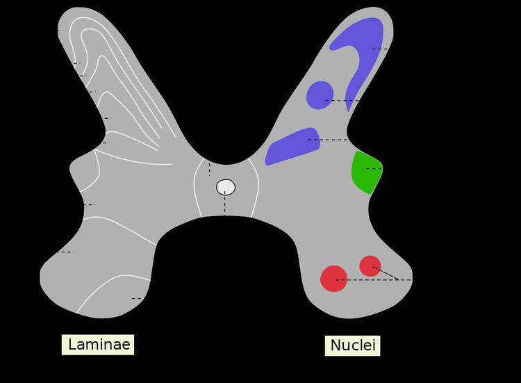
The Rexed laminae comprise a system of ten layers of grey matter (I-X), identified in the early 1950s by Bror Rexed to label portions of the grey columns of the spinal cord.

Similar to Brodmann areas, they are defined by their cellular structure rather than by their location, but the location still remains reasonably consistent.

Laminae

  • Posterior/dorsal horn: I-VI
  • Lamina I: marginal nucleus of spinal cord or posteromarginal nucleus
  • Lamina II: substantia gelatinosa of Rolando
  • Laminae III and IV: nucleus proprius
  • Lamina V: Neck of the dorsal horn. Neurons within lamina V are mainly involved in processing sensory afferent stimuli from cutaneous, muscle and joint mechanical nociceptors as well as visceral nociceptors. This layer is home to wide dynamic range tract neurons, interneurons and propriospinal neurons. Viscerosomatic pain signal convergence often occurs in this lamina due to the presence of wide dynamic range tract neurons resulting in pain referral.
  • Lamina VI: Base of the dorsal horn. 'Fast pain' moves to lamina VI, which controls flexion reflex (i.e. withdrawal from painful stimulus).
  • Intermediate zone: VII and X
  • Lamina VII: intermediomedial nucleus, intermediolateral nucleus, posterior thoracic nucleus in the thoracic and upper lumbar region
  • Lamina X: central gray matter i.e. neurons bordering Central canal; Grisea Centralis
  • Anterior/ventral horn: VIII-IX
  • Lamina VIII: motor interneurons; Commissural nucleus
  • Lamina IX: lateral (in limb regions) and medial motor neurons, also phrenic and spinal accessory nuclei at cervical levels, and Onuf's nucleus in the sacral region
  • Centrally
  • Lamina X: Central Zone, grey matter surrounding the central canal
  • References

    Rexed laminae Wikipedia