
MedlinePlus 000266 MeSH C23.300.825 | ICD-10 K63.5 & various eMedicine med/414 | |
 | ||
A polyp is an abnormal growth of tissue projecting from a mucous membrane. If it is attached to the surface by a narrow elongated stalk, it is said to be pedunculated. If no stalk is present, it is said to be sessile. Polyps are commonly found in the colon, stomach, nose, ear, sinus(es), urinary bladder, and uterus. They may also occur elsewhere in the body where mucous membranes exist like the cervix, vocal folds, and small intestine. Some polyps are tumors (neoplasms) and others are nonneoplastic (for example, hyperplastic or dysplastic). The neoplastic ones are generally benign, although some can be premalignant and/or concurrent with a malignancy.
Contents
Localization
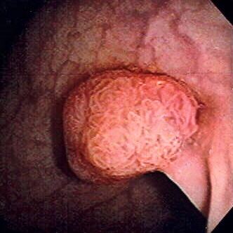
Colorectal polyp
Colon polyps are not commonly associated with symptoms. Occasionally rectal bleeding, and on rare occasions pain, diarrhea or constipation may occur because of colon polyps. Colon polyps are a concern because of the potential for colon cancer being present microscopically and the risk of benign colon polyps transforming over time into malignant ones. Since most polyps are asymptomatic, they are usually discovered at the time of colon cancer screening. Common screening methods are occult blood test, colonoscopy, sigmoidoscopy (usually flexible sigmoidoscopy, using a flexible endoscope, but more rarely the older rigid sigmoidoscopy, using a rigid endoscope), lower gastrointestinal series (barium enema), digital rectal examination (DRE), and virtual colonoscopy. The polyps are routinely removed at the time of colonoscopy either with a polypectomy snare (first description by P. Deyhle, Germany, 1970) or with biopsy forceps. If an adenomatous polyp is found with sigmoidoscopy or if a polyp is found with any other diagnostic modality, the patient must undergo colonoscopy for removal of the polyp(s). Even though colon cancer is usually not found in polyps smaller than 2.5 cm, all polyps found are removed since the removal of polyps reduces the future likelihood of developing colon cancer. When adenomatous polyps are removed, a repeat colonoscopy is usually performed in three to five years.
Most colon polyps can be categorized as sporadic.
Inherited polyposis syndromes
Non-inherited polyposis syndromes
Types of colon polyps
Adenomatous polyps
Adenomatous polyps, or adenomas, are polyps that grow on the lining of the colon and which carry a high risk of cancer. The adenomatous polyp is considered pre-malignant, i.e., likely to develop into colon cancer. The other types of polyps that can occur in the colon are the hyperplastic and inflammatory polyps. They are unlikely to develop into colorectal cancer.
About 5% of people aged 60 will have at least one adenomatous polyp of 1 cm diameter or greater. Multiple adenomatous polyps often result from familial polyposis coli or familial adenomatous polyposis, a condition that carries a very high risk of colon cancer.
Types
Adenomas constitute approximately 10% of digestive polyps. Most polyps (approximately 90%) are small, usually less than 1 cm in diameter, and have a small potential for malignancy. The remaining 10% of adenomas are larger than 1 cm and approach a 10% chance of containing invasive cancer.
There are three types of adenomatous polyp:
Risks
The risks of progression to colorectal cancer increases if the polyp is larger than 1 cm and contains a higher percentage of villous component. Also, the shape of the polyps is related to the risk of progression into carcinoma. Polyps that are pedunculated (with a stalk) are usually less dangerous than sessile polyps (flat polyps). Sessile polyps have a shorter pathway for migration of invasive cells from the tumor into submucosal and more distant structures, and they are also more difficult to remove and to ascertain. Sessile polyps larger than 2 cm usually contain villous features, have a higher malignant potential, and tend to recur following colonoscopic polypectomy.
Although polyps do not carry significant risk of colon cancer, tubular adenomatous polyps may become cancerous when they grow larger. Larger tubular adenomatous polyps have an increased risk of malignancy when larger because then they develop more villous components and may become sessile.
It is estimated that an individual whose parents have been diagnosed with an adenomatous polyp has a 50% greater chance to develop colon cancer than individuals with no family history of colonic polyps. At this point, there is no method to establish the risks that patients with a family history of colon polyps have to develop these growths. Overall, nearly 6% of the population, regardless of the family history, is at risk of developing colon cancer.
Screening
Screening for colonic polyps as well as preventing them has become an important part of the management of the condition. Medical societies have established guidelines for colorectal screening in order to prevent adenomatous polyps and to minimize the chances of developing colon cancer. It is believed that some changes in the diet might be helpful in preventing polyps from occurring but there is no other way to prevent the polyps from developing into cancerous growths than by detecting and removing them.
According to the guidelines established by the American Cancer Society, individuals who reach the age of 50 should perform an occult blood test yearly. Colon polyps as they grow can sometimes cause bleeding within the intestine, which can be detected with the help of this test. Also, persons in their 50s are recommended to have flexible sigmoidoscopies performed once in 3 to 5 years to detect any abnormal growth which could be an adenomatous polyp. If adenomatous polyps are detected during this procedure, it is most likely that the patient will have to undergo a colonoscopy. Medical societies recommend colonoscopies every ten years starting at age 50 as a necessary screening practice for colon cancer. The screening provides an accurate image of the intestine and also allows the removal of the polyp, if found. Once an adenomatous polyp is identified during colonoscopy, there are several methods of removal including using a snare or a heating device. Colonoscopies are preferred over sigmoidoscopies because they allow the examination of the entire colon; a very important aspect, considering that more than half of the colonic polyps occur in the upper colon, which is not reached during sigmoidoscopies.
It has been statistically demonstrated that screening programs are effective in reducing the number of deaths caused by colon cancer due to adenomatous polyps. While there are risks of complications associated with colonoscopies, those risks are extremely low at approximately 0.35 percent. For comparison, the lifetime risk of developing colon cancer is around 6 percent. As there is a small likelihood of recurrence, surveillance after polyp removal is recommended.
Endometrial polyp
An endometrial polyp or uterine polyp is a polyp or lesion in the lining of the uterus (endometrium) that takes up space within the uterine cavity. Commonly occurring, they are experienced by up to 10% of women. They may have a large flat base (sessile) or be attached to the uterus by an elongated pedicle (pedunculated). Pedunculated polyps are more common than sessile ones. They range in size from a few millimeters to several centimeters. If pedunculated, they can protrude through the cervix into the vagina. Small blood vessels may be present in polyps, particularly large ones.
Cervical polyp
A cervical polyp is a common benign polyp or tumor on the surface of the cervical canal. They can cause irregular menstrual bleeding or increased pain but often show no symptoms.
Nasal polyps
Nasal polyps are polypoidal masses arising mainly from the mucous membranes of the nose and paranasal sinuses. They are overgrowths of the mucosa that frequently accompany allergic rhinitis. They are freely movable and nontender.
Laryngeal polyps
Polyps on the vocal folds can take on many different forms, and can sometimes result from vocal abuse, although this is not always the cause. They can occur on one or both vocal folds, and appear as swelling, a bump (similar to a nodule), a stalk-like growth, or a blister-like lesion. Most polyps are larger than nodules, which are more similar to callouses on the vocal folds.
Polyps and nodules can exhibit similar symptoms including hoarseness or breathiness, “rough” or “scratchy” voice, harshness in vocal quality, shooting pain from ear to ear, sensation of having “a lump in the back of the throat”, neck pain, decreased pitch range in the voice, and vocal and bodily fatigue.
If an individual experiences symptoms for more than 2 to 3 weeks, they should see a physician. For a diagnosis, a thorough evaluation of the voice should include a physical examination, preferably by an otolaryngologist (ear, nose, and throat doctor) who specializes in voice, a voice evaluation with a speech-language pathologist (SLP), a neurological examination (in certain cases) The qualities of the voice that will be evaluated include quality, pitch, loudness, and ability to sustain voicing. In some cases, an instrumental examination may be performed with an endoscope into the mouth or nose; this gives a clear look at the vocal folds and larynx in general. In addition to this, a stroboscope (flashing light) may be used to observe the movement of the vocal folds during speech.
Polyps may be treated with medical, surgical, or behavioral intervention. Surgical intervention involves removing the polyp from the vocal fold. This approach is only used when the growth(s) are very large, or have existed for an extended amount of time. In children, surgical intervention is rare. Existing medical problems may be treated in an effort to reduce the strain and negative impact on the vocal cords. This could include treatment for gastrointestinal reflux disease, allergies, and thyroid problems. Intervention to stop smoking and reduce stress may also be needed. Most people receive behavioral intervention, or vocal therapy, from an SLP. This might involve teaching good vocal hygiene, and reducing or stopping vocal abuse behaviors. Direct voice treatments may be used to alter pitch, loudness, or breathe support to promote good voicing.