
EC number 1.6.5.3 BRENDA BRENDA entry KEGG KEGG entry | IntEnz IntEnz view ExPASy NiceZyme view MetaCyc | |
 | ||
Complex I (EC 1.6.5.3) (also referred to as NADH:ubiquinone oxidoreductase or, especially in the context of the human protein, NADH dehydrogenase) is an enzyme of the respiratory chains of myriad organisms from bacteria to humans that falls under the H+ or Na+-translocating NADH Dehydrogenase (NDH) Family (TC# 3.D.1), a member of the Na+ transporting Mrp superfamily. It catalyzes the transfer of electrons from NADH to coenzyme Q10 (CoQ10) and, in eukaryotes, it is located in the inner mitochondrial membrane. NADH:ubiquinone oxidoreductases type I of bacteria and of eukaryotic mitochondria and chloroplasts couple electron transfer to the electrogenic transport of protons or Na+. It is one of the "entry enzymes" of cellular respiration or oxidative phosphorylation in the mitochondria.
Contents
Function
Complex I is the first enzyme of the mitochondrial electron transport chain. There are three energy-transducing enzymes in the electron transport chain - NADH:ubiquinone oxidoreductase (complex I), Coenzyme Q – cytochrome c reductase (complex III), and cytochrome c oxidase (complex IV). Complex I is the largest and most complicated enzyme of the electron transport chain.
The reaction catalyzed by complex I is:
NADH + H+ + CoQ + 4H+in→ NAD+ + CoQH2 + 4H+outIn this process, the complex translocates four protons across the inner membrane per molecule of oxidized NADH, helping to build the electrochemical potential difference used to produce ATP. Escherichia coli complex I (NADH dehydrogenase) is capable of proton translocation in the same direction to the established Δψ, showing that in the tested conditions, the coupling ion is H+. Na+ transport in the opposite direction was observed, and although Na+ was not necessary for the catalytic or proton transport activities, its presence increased the latter. H+ was translocated by the Paracoccus denitrificans complex I, but in this case, H+ transport was not influenced by Na+, and Na+ transport was not observed. Possibly, the E. coli complex I has two energy coupling sites (one Na+ independent and the other Na+dependent), as observed for the Rhodothermus marinus complex I, whereas the coupling mechanism of the P. denitrificans enzyme is completely Na+ independent. It is also possible that another transporter catalyzes the uptake of Na+. Complex I energy transduction by proton pumping may not be exclusive to the R. marinus enzyme. The Na+/H+ antiport activity seems not to be a general property of complex I. However, the existence of Na+-translocating activity of the complex I is still in question.
The reaction can be reversed – referred to as aerobic succinate-supported NAD+ reduction by ubiquinol – in the presence of a high membrane potential, but the exact catalytic mechanism remains unknown. Driving force of this reaction is a potential across the membrane which can be maintained eitehr by ATP-hydrolysis or by complexes III and IV during succinate oxidation.
Complex I may have a role in triggering apoptosis. In fact, there has been shown to be a correlation between mitochondrial activities and programmed cell death (PCD) during somatic embryo development.
Overall Mechanism
All redox reactions take place in the hydrophilic domain of complex I. NADH initially binds to complex I, and transfers two electrons to the flavin mononucleotide (FMN) prosthetic group of the enzyme, creating FMNH2. The electron acceptor – the isoalloxazine ring – of FMN is identical to that of FAD. The electrons are then transferred through the FMN via a series of iron-sulfur (Fe-S) clusters, and finally to coenzyme Q10 (ubiquinone). This electron flow changes the redox state of the protein, inducing conformational changes of the protein which alters the pK values of ionizable side chain, and causes four hydrogen ions to be pumped out of the mitochondrial matrix. Ubiquinone (CoQ) accepts two electrons to be reduced to ubiquinol (CoQH2).
Electron Transfer Mechanism
The proposed pathway for electron transport prior to ubiquinone reduction is as follows: NADH – FMN – N3 – N1b – N4 – N5 – N6a – N6b – N2 – Q, where Nx is a labelling convention for iron sulfur clusters. The high reduction potential of the N2 cluster and the relative proximity of the other clusters in the chain enable efficient electron transfer over long distance in the protein (with transfer rates from NADH to N2 iron-sulfur cluster of about 100 μs).
The equilibrium dynamics of Complex I are primarily driven by the quinone redox cycle. In conditions of high proton motive force (and accordingly, a ubiquinol-concentrated pool), the enzyme runs in the reverse direction. Ubiquinol is oxidized to ubiquinone, and the resulting released protons reduce the proton motive force.
Proton Translocation Mechanism
The coupling of proton translocation and electron transport in Complex I is currently proposed as being indirect (long range conformational changes) as opposed to direct (redox intermediates in the hydrogen pumps as in heme groups of Complexes III and IV). The architecture of the hydrophobic region of complex I shows multiple proton transporters that are mechanically interlinked. The three central components believed to contribute to this long-range conformational change event are the pH-coupled N2 iron-sulfur cluster, the quinone reduction, and the transmembrane helix subunits of the membrane arm. Transduction of conformational changes to drive the transmembrane transporters linked by a 'connecting rod' during the reduction of ubiquinone can account for two or three of the four protons pumped per NADH oxidized. The remaining proton must be pumped by direct coupling at the ubiquinone-binding site. It is proposed that direct and indirect coupling mechanisms account for the pumping of the four protons.
The N2 cluster's proximity to a nearby cysteine residue results in a conformational change upon reduction in the nearby heliceses, leading to small but important changes in the overall protein conformation. Further electron paramagnetic resonance studies of the electron transfer have demonstrated that most of the energy that is released during the subsequent CoQ reduction is on the final ubiquinol formation step from semiquinone, providing evidence for the "single stroke" H+ translocation mechanism (i.e. all four protons move across the membrane at the same time). Alternative theories suggest a "two stroke mechanism" where each reduction step (semiquinone and ubiquinol) results in a stroke of two protons entering the intermembrane space.
The resulting ubiquinol localized to the membrane domain interacts with negatively charged residues in the membrane arm, stabilizing conformational changes. An antiporter mechanism (Na+/H+ swap) has been proposed using evidence of conserved Asp residues in the membrane arm. The presence of Lys, Glu, and His residues enable for proton gating (a protonation followed by deprotonation event across the membrane) driven by the pKa of the residues.
Composition and structure
NADH:ubiquinone oxidoreductase is the largest of the respiratory complexes. In mammals, the enzyme contains 44 separate water soluble peripheral membrane proteins, which are anchored to the integral membrane constituents. Of particular functional importance are the flavin prosthetic group (FMN) and eight iron-sulfur clusters (FeS). Of the 44 subunits, seven are encoded by the mitochondrial genome.
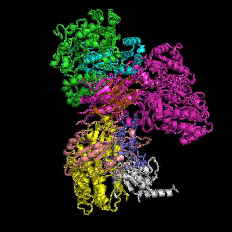
The structure is an "L" shape with a long membrane domain (with around 60 trans-membrane helices) and a hydrophilic (or peripheral) domain, which includes all the known redox centres and the NADH binding site. The structure of the eukaryotic complex is not well characterised. However, the Sazanov group succeeded in solving the structures of the complex I hydrophilic domain from the bacterium Thermus thermophilus (PDB: 2FUG) and complex I membrane domains from both the E. coli (PDB: 3rko) and T. thermophilus (PDB: 4HE8) enzymes. In February 2013 the structure of an entire, intact complex I (from T. thermophilus) was published for the first time, again by the Sazanov group (PDB: 4HEA). All thirteen of the E. coli proteins, which comprise NADH dehydrogenase I, are encoded within the nuo operon, and are homologous to mitochondrial complex I subunits. The antiporter-like subunits NuoL/M/N each contains 14 conserved transmembrane (TM) helices. Two of them are discontinuous, but subunit NuoL contains a 110 Å long amphipathic α-helix, spanning the entire length of the domain. The subunit, NuoL, is related to Na+/ H+ antiporters of TC# 2.A.63.1.1 (PhaA and PhaD).
Three of the conserved, membrane-bound subunits in NADH dehydrogenase are related to each other, and to Mrp sodium-proton antiporters. Structural analysis of two prokaryotic complexes I revealed that the three subunits each contain fourteen transmembrane helices that overlay in structural alignments: the translocation of three protons may be coordinated by a lateral helix connecting them.
Complex I contains a ubiquinone binding pocket at the interface of the 49-kDa and PSST subunits. Close to iron-sulfur cluster N2, the proposed immediate electron donor for ubiquinone, a highly conserved tyrosine constitutes a critical element of the quinone reduction site. A possible quinone exchange path leads from cluster N2 to the N-terminal beta-sheet of the 49-kDa subunit. All 45 subunits of the bovine NDHI have been sequenced. Each complex contains noncovalently bound FMN, coenzyme Q and several iron-sulfur centers. The bacterial NDHs have 8-9 iron-sulfur centers.
A recent study by Roessler et al. (2010) used electron paramagnetic resonance (EPR) spectra and double electron-electron resonance (DEER) to determine the path of electron transfer through the iron-sulfur complexes, which are located in the hydrophilic domain. Seven of these clusters form a chain from the flavin to the quinone binding sites; the eighth cluster is located on the other side of the flavin, and its function is unknown. The EPR and DEER results suggest an alternating or “roller-coaster” potential energy profile for the electron transfer between the active sites and along the iron-sulfur clusters, which can optimize the rate of electron travel and allow efficient energy conversion in complex I.
A simulational study by Hayashi and Stuchebrukhov further identified the electron tunneling pathways in atomic resolution based on the tunneling current theory. The distinct pathways between neighboring Fe/S clusters primarily consist of two cysteine ligands and one additional key residue, which was supported by sensitivity of simulated electron transfer rates to their mutations and their conservation among various complex I homologues from simple bacteria to human beings. This result shows that the crucial part of complex I developed for optimal efficiency with specific key residues during early stages of the biological evolution and has been conserved since then. Internal water between protein subunits was identified as an essential mediator enhancing the overall electron transfer rate to achieve physiologically significant value.
Table of Conserved subunits of Complex I
Inhibitors
The best-known inhibitor of complex I is rotenone (commonly used as an organic pesticide). Rotenone and rotenoids are isoflavonoids occurring in several genera of tropical plants such as Antonia (Loganiaceae), Derris and Lonchocarpus (Faboideae, Fabaceae). There have been reports of the indigenous people of French Guiana using rotenone-containing plants to fish - due to its ichthyotoxic effect - as early as the 17th century. Rotenone binds to the ubiquinone binding site of complex I as well as piericidin A, another potent inhibitor with a close structural homologue to ubiquinone.
Acetogenins from Annonaceae are even more potent inhibitors of complex I. They cross-link to the ND2 subunit, which suggests that ND2 is essential for quinone-binding. Interestingly, Rolliniastatin-2, an acetogenin, is the first complex I inhibitor found that does not share the same binding site as rotenone.
Despite more than 50 years of study of complex I, no inhibitors blocking the electron flow inside the enzyme have been found. Hydrophobic inhibitors like rotenone or piericidin most likely disrupt the electron transfer between the terminal FeS cluster N2 and ubiquinone. It has been shown that long-term systemic inhibition of complex I by rotenone can induce selective degeneration of dopaminergic neurons.
Complex I is also blocked by adenosine diphosphate ribose – a reversible competitive inhibitor of NADH oxidation – by binding to the enzyme at the nucleotide binding site. Both hydrophilic NADH and hydrophobic ubiquinone analogs act at the beginning and the end of the internal electron-transport pathway, respectively.
The antidiabetic drug Metformin has been shown to induce a mild and transient inhibition of the mitochondrial respiratory chain complex I, and this inhibition appears to play a key role in its mechanism of action.
Inhibition of complex I has been implicated in hepatotoxicity associated with a variety of drugs, for instance flutamide and nefazodone.
Active/deactive transition
The catalytic properties of eukaryotic complex I are not simple. Two catalytically and structurally distinct forms exist in any given preparation of the enzyme: one is the fully competent, so-called “active” A-form and the other is the catalytically silent, dormant, “deactive”, D-form. After exposure of idle enzyme to elevated, but physiological temperatures (>30 °C) in the absence of substrate, the enzyme converts to the D-form. This form is catalytically incompetent but can be activated by the slow reaction (k~4 min−1) of NADH oxidation with subsequent ubiquinone reduction. After one or several turnovers the enzyme becomes active and can catalyse physiological NADH:ubiquinone reaction at a much higher rate (k~104 min−1). In the presence of divalent cations (Mg2+, Ca2+), or at alkaline pH the activation takes much longer.
The high activation energy (270 kJ/mol) of the deactivation process indicates the occurrence of major conformational changes in the organisation of the complex I. However, until now, the only conformational difference observed between these two forms is the number of cysteine residues exposed at the surface of the enzyme. Treatment of the D-form of complex I with the sulfhydryl reagents N-Ethylmaleimide or DTNB irreversibly blocks critical cysteine residue(s), abolishing the ability of the enzyme to respond to activation, thus inactivating it irreversibly. The A-form of complex I is insensitive to sulfhydryl reagents.
It was found that these conformational changes may have a very important physiological significance. The deactive, but not the active form of complex I was susceptible to inhibition by nitrosothiols and peroxynitrite. It is likely that transition from the active to the inactive form of complex I takes place during pathological conditions when the turnover of the enzyme is limited at physiological temperatures, such as during hypoxia, or when the tissue nitric oxide:oxygen ratio increases (i.e. metabolic hypoxia).
Production of superoxide
Recent investigations suggest that complex I is a potent source of reactive oxygen species. Complex I can produce superoxide (as well as hydrogen peroxide), through at least two different pathways. During forward electron transfer, only very small amounts of superoxide are produced (probably less than 0.1% of the overall electron flow).
During reverse electron transfer, complex I might be the most important site of superoxide production within mitochondria, with up to 5% of electrons being diverted to superoxide formation. Reverse electron transfer, the process by which electrons from the reduced ubiquinol pool (supplied by succinate dehydrogenase, glycerol-3-phosphate dehydrogenase, or dihydro-oorotate dehydrogenase in mammalian mitochondria) pass through complex I to reduce NAD+ to NADH, driven by the inner mitochondrial membrane potential electric potential. Although it is not precisely known under what pathological conditions reverse-electron transfer would occur in vivo, in vitro experiments indicate that it can be a very potent source of superoxide when succinate concentrations are high and oxaloacetate or malate concentrations are low.
Superoxide is a reactive oxygen species that contributes to cellular oxidative stress and is linked to neuromuscular diseases and aging. NADH dehdyrogenase produces superoxide by transferring one electron from FMNH2 to oxygen (O2). The radical flavin leftover is unstable, and transfers the remaining electron to the iron-sulfur centers. Interestingly, it is the ratio of NADH to NAD+ that determines the rate of superoxide formation.
Pathology
Mutations in the subunits of complex I can cause mitochondrial diseases, including Leigh syndrome. Point mutations in various complex I subunits derived from mitochondrial DNA (mtDNA) can also result in Leber's Hereditary Optic Neuropathy. There is some evidence that complex I defects may play a role in the etiology of Parkinson's disease, perhaps because of reactive oxygen species (complex I can, like complex III, leak electrons to oxygen, forming highly toxic superoxide).
Although the exact etiology of Parkinson’s disease is unclear, it is likely that mitochondrial dysfunction, along with proteasome inhibition and environmental toxins, may play a large role. In fact, the inhibition of complex I has been shown to cause the production of peroxides and a decrease in proteasome activity, which may lead to Parkinson’s disease. Additionally, Esteves et al. (2010) found that cell lines with Parkinson’s disease show increased proton leakage in complex I, which causes decreased maximum respiratory capacity.
Recent studies have examined other roles of complex I activity in the brain. Andreazza et al. (2010) found that the level of complex I activity was significantly decreased in patients with bipolar disorder, but not in patients with depression or schizophrenia. They found that patients with bipolar disorder showed increased protein oxidation and nitration in their prefrontal cortex. These results suggest that future studies should target complex I for potential therapeutic studies for bipolar disorder. Similarly, Moran et al. (2010) found that patients with severe complex I deficiency showed decreased oxygen consumption rates and slower growth rates. However, they found that mutations in different genes in complex I lead to different phenotypes, thereby explaining the variations of pathophysiological manifestations of complex I deficiency.
Exposure to pesticides can also inhibit complex I and cause disease symptoms. For example, chronic exposure to low levels of dichlorvos, an organophosphate used as a pesticide, has been shown to cause liver dysfunction. This occurs because dichlorvos alters complex I and II activity levels, which leads to decreased mitochondrial electron transfer activities and decreased ATP synthesis.
Genes
The following is a list of humans genes that encode components of complex I: