
 | ||
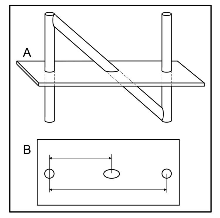
The N-localizer (aka N-bar) is a device that enables guidance of stereotactic surgery or radiosurgery using tomographic images that are obtained via medical imaging technologies such as X-ray computed tomography (CT), magnetic resonance imaging (MRI) or positron emission tomography (PET). The N-localizer comprises a diagonal rod that extends from the top of one vertical rod to the bottom of a second vertical rod, and is shaped like a capital "N" (Figure 1A). This device was invented in 1978 by the American physician and computer scientist Russell Brown.
This invention stimulated renewed interest in, and further development of, image-guided surgery, specifically, stereotactic surgery and radiosurgery. It has achieved widespread clinical use in the Brown-Roberts-Wells (BRW), Leksell, Kelly-Goerss and Cosman-Roberts-Wells (CRW) stereotactic instruments and in the Gamma Knife streotactic radiosurgery system.
How it works
Medical images that are obtained using technologies such as computed tomography (CT), magnetic resonance imaging (MRI) and positron emission tomography (PET) are two-dimensional, planar, tomographic images of patient anatomy. Treatment modalities such as stereotactic surgery and radiosurgery operate in the three-dimensional space of the patient. Hence, the central problem with using CT, MRI or PET to guide stereotactic surgery or radiosurgery is the transfer of patient information from the two-dimensional coordinate system of the planar CT, MRI or PET image into the three-dimensional coordinate system of the stereotactic or radiosurgical instrument. The N-localizer provides an elegant solution to this problem by creating fiducial marks or landmarks in each tomographic image. The N-localizer comprises a diagonal rod that extends from the top of one vertical rod to the bottom of a second vertical rod (Figure 1). This combination of rods creates two circles and one ellipse in a tomographic image. The ellipse moves away from one circle and towards the other circle as the image plane moves downward with respect to the N-localizer. Measuring the relative distances between the ellipse and the two circles permits calculation of the point where the image plane intersects the diagonal rod. The attachment of three N-localizers to a stereotactic instrument (Figure 2) permits calculation of three points where the image plane intersects the three diagonal rods (Figure 3). Because three points determine a plane in three-dimensional space, these three points of intersection determine the spatial orientation of the image plane relative to the stereotactic instrument. Therefore, the spatial orientation of any patient anatomy that is seen in the planar image is also determined relative to the stereotactic instrument. Because the spatial orientation of the patient anatomy is determined unambiguously relative to the stereotactic instrument, patient information may be transferred from the two-dimensional coordinate system of the planar image into the three-dimensional coordinate system of the stereotactic instrument.