
Group Group I (dsDNA) Family Herpesviridae | Order Herpesvirales Subfamily Alphaherpesvirinae | |
 | ||
Species Gallid herpesvirus 2 (GaHV-2) | ||
Marek's disease is a highly contagious viral neoplastic disease in chickens. It is named after József Marek, a Hungarian veterinarian. Occasionally misdiagnosed as an abtissue pathology it is caused by an alphaherpesvirus known as 'Marek's disease virus' (MDV) or Gallid herpesvirus 2 (GaHV-2). The disease is characterized by the presence of T cell lymphoma as well as infiltration of nerves and organs by lymphocytes. Viruses related to MDV appear to be benign and can be used as vaccine strains to prevent Marek's disease. For example, the related Herpesvirus of Turkeys (HVT), causes no apparent disease in turkeys and continues to be used as a vaccine strain for prevention of Marek's disease (see below). Birds infected with GaHV-2 can be carriers and shedders of the virus for life. Newborn chicks are protected by maternal antibodies for a few weeks. After infection, microscopic lesions are present after one to two weeks, and gross lesions are present after three to four weeks. The virus is spread in dander from feather follicles and transmitted by inhalation.
Contents
Syndromes
Six syndromes are known to occur after infection with Marek's disease. These syndromes may overlap.
Diagnosis
Diagnosis of lymphoid tumors in poultry is complicated due to multiple etiological agents capable of causing very similar tumors. It is not uncommon that more than one avian tumor virus can be present in a chicken, thus one must consider both the diagnosis of the disease/tumors (pathological diagnosis) and of the virus (etiological diagnosis). A step-wise process has been proposed for diagnosis of Marek’s disease which includes (1) history, epidemiology, clinical observations and gross necropsy, (2) characteristics of the tumor cell, and (3) virological characteristics
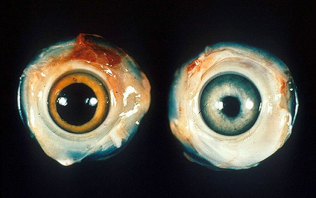
The demonstration of peripheral nerve enlargement along with suggestive clinical signs in a bird that is around three to four months old (with or without visceral tumors) is highly suggestive of Marek's disease. Histological examination of nerves reveals infiltration of pleomorphic neoplastic and inflammatory lymphocytes. Peripheral neuropathy should also be considered as a principal rule-out in young chickens with paralysis and nerve enlargement without visceral tumors, especially in nerves with interneuritic edema and infiltration of plasma cells.
The presence of nodules on the internal organs may also suggest Marek's disease, but further testing is required for confirmation. This is done through histological demonstration of lymphomatous infiltration into the affected tissue. A range of leukocytes can be involved, including lymphocytic cell lines such as large lymphocyte, lymphoblast, primitive reticular cells, and occasional plasma cells, as well as macrophage and plasma cells. The T cells are involved in the malignancy, showing neoplastic changes with evidence of mitosis. The lymphomatous infiltrates need to be differentiated from other conditions that affect poultry including lymphoid leukosis and reticuloendotheliosis, as well as an inflammatory event associated with hyperplastic changes of the affected tissue.
Key clinical signs as well as gross and microscopic features that are most useful for differentiating Marek’s disease from lymphoid leukosis and reticuloendotheliosis include (1) Age: MD can affect birds at any age, including <16 weeks of age; (2) Clinical signs: Frequent wing and leg paralysis; (3) Incidence: >5% in unvaccinated flocks; (4) Potential nerve enlargement; (5) Interfollicular tumors in the bursa of Fabricius; (6) CNS involvement; (7) Lymphoid proliferation in skin and feather follicles; (8) Pleomorphic lymphoid cells in nerves and tumors; and (9) T-cell lymphomas.
In addition to gross pathology and histology, other advanced procedures used for a definitive diagnosis of Marek’s disease include immunohistochemistry to identify cell type and virus-specific antigens, standard and quantitative PCR for identification of the virus, virus isolation to confirm infections, and serology to confirm/exclude infections.
The World Organisation for Animal Health (OIE) reference laboratories for Marek’s disease include the Institute for Animal Health, Compton Laboratory, UK and the USDA Avian Disease and Oncology Laboratory, USA.
Prevention
Vaccination is the only known method to prevent the development of tumors when chickens are infected with the virus. However, administration of vaccines does not prevent transmission of the virus, i.e., the vaccine is not sterilizing. However, it does reduce the amount of virus shed in the dander, hence reduces horizontal spread of the disease. Marek's disease does not spread vertically. The vaccine was introduced in 1970 and the scientist credited with its development is Dr. Ben Roy Burmester. Before that, Marek's disease caused substantial revenue loss in the poultry industries of the United States and the United Kingdom. The vaccine can be administered to one-day-old chicks through subcutaneous inoculation or by in ovo vaccination when the eggs are transferred from the incubator to the hatcher. In ovo vaccination is the preferred method, as it does not require handling of the chicks and can be done rapidly by automated methods. Immunity develops within two weeks.
The vaccine originally contained the antigenically similar turkey herpesvirus, which is serotype 3 of MDV. However, because vaccination does not prevent infection with the virus, the Marek's disease virus has evolved increased virulence and resistance to this vaccine. As a result, current vaccines use a combination of vaccines consisting of HVT and gallid herpesvirus type 3 or an attenuated MDV strain, CVI988-Rispens (ATCvet code: QI01AD03 (WHO)).