
Latin vas lymphocapillare TA A12.0.00.044 | Code TH H3.09.02.0.05004 FMA 5028 | |
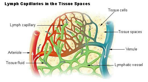 | ||
Lymph capillaries or lymphatic capillaries are tiny, thin-walled vessels located in the spaces between cells (except in the central nervous system and non-vascular tissues) which serve to drain and process extra-cellular fluid. Upon entering the lumen of a lymphatic capillary, the collected fluid and associated cells (notably white blood cells) is known as lymph. Each lymphatic capillary carries lymph into a lymphatic vessel, which in turn connects to a lymph node. Lymph is ultimately returned to the venous circulation.
Lymphatic capillaries are slightly larger in diameter than blood capillaries, and have closed ends (unlike the loop structure of blood capillaries). Their unique structure permits interstitial fluid to flow into them but not out. The ends of the endothelial cells that make up the wall of a lymphatic capillary overlap. When pressure is greater in the interstitial fluid than in lymph, the cells separate slightly, like the opening of a one-way swinging door, and interstitial fluid enters the lymphatic capillary. When pressure is greater inside the lymphatic capillary, the cells adhere more closely, and lymph cannot escape back into interstitial fluid. Attached to the lymphatic capillaries are anchoring filaments, which contain elastic fibers. They extend out from the lymphatic capillary, attaching lymphatic endothelial cells to surrounding tissues. When excess interstitial fluid accumulates and causes tissue swelling, the anchoring filaments are pulled, making the openings between cells even larger so that more fluid can flow into the lymphatic capillary.
Lymph capillaries have a greater internal oncotic pressure than blood capillaries, due to the greater concentration of plasma proteins in the lymph.