
 | ||
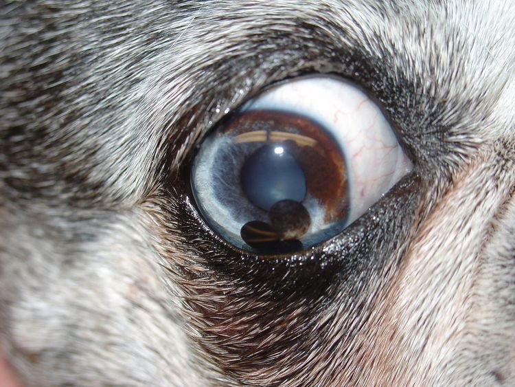
An iris cyst, or uveal cyst, is a small hollow structure either attached to the iris of the eye or floating free in the anterior chamber. An iris cyst is composed of a single cell layer of epithelium and is filled with fluid. It is most commonly seen as secondary to inflammation in the eye, especially with canine glaucoma. They are most commonly seen in dogs. Golden Retrievers, Labrador Retrievers, and Boston Terriers are the most commonly affected breeds. Iris cysts also occur in cats and horses. The cysts are usually free floating in dogs, attached to the pupillary margin in cats, and present in the interior of the iris (especially blue irises) in horses.
Iris cysts usually cause no symptoms, but in large numbers they can cause glaucoma by obstructing the drainage angle of the eye. Golden Retrievers may have a higher rate of glaucoma associated with iris cysts.
Iris cysts need to be differentiated from iris melanoma, a tumor, by demonstrating their hollowness. A light directed at an iris cyst will cause it to transilluminate. Iris cysts can be collapsed if necessary by a veterinarian with a small needle carefully introduced into the anterior chamber. The cysts may also be destroyed by use of a semiconductor diode laser.