
Dorlands
/Elsevier p_24/12648710 | FMA 4881 | |
 | ||
Latin plexus venosi vertebrales interni TA A12.3.07.021
A12.3.07.026 | ||
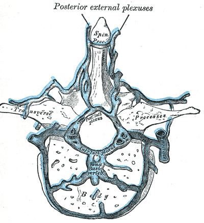
The internal vertebral venous plexuses (intraspinal veins) lie within the vertebral canal in the epidural space, and receive tributaries from the bones and from the medulla spinalis.
They form a closer network than the external plexuses, and, running mainly in a vertical direction, form four longitudinal veins, two in front and two behind; they therefore may be divided into anterior and posterior groups.
The anterior and posterior plexuses communicate freely with one another by a series of venous rings (retia venosa vertebrarum), one opposite each vertebra.
Around the foramen magnum they form an intricate network which opens into the vertebral veins and is connected above with the occipital sinus, the basilar plexus, the condyloid emissary vein, and the rete canalis hypoglossi.