
 | ||
In cell biology, ways in which fragmentation is useful for a cell: DNA cloning and apoptosis. DNA cloning is important in asexual reproduction or creation of identical DNA molecules, and can be performed spontaneously by the cell or intentionally by laboratory researchers. Apoptosis is the programmed destruction of cells, and the DNA molecules within them, and is a highly regulated process. These two ways in which fragmentation is used in cellular processes describe normal cellular functions and common laboratory procedures performed with cells. However, problems within a cell can sometimes cause fragmentation that results in irregularities such as red blood cell fragmentation and sperm cell DNA fragmentation.
Contents
DNA Cloning
DNA cloning can be performed spontaneously by the cell for reproductive purposes. This is a form of asexual reproduction where an organism splits into fragments and then each of these fragments develop into mature, fully grown individuals that are clones of the original organism (See reproductive fragmentation). DNA cloning can also be performed intentionally by laboratory researchers. Here, DNA fragmentation is a molecular genetic technique that permits researchers to use recombinant DNA technology to prepare large numbers of identical DNA molecules. In order for DNA cloning to be completed, it is necessary to obtain discrete, small regions of an organism's DNA that constitute specific genes. Only relatively small DNA molecules can be cloned in any available vector. Therefore, the long DNA molecules that compose an organism's genome must be cleaved into fragments that can be inserted into the vector DNA. Two enzymes facilitate the production of such recombinant DNA molecules:
1. Restriction Enzymes2. DNA ligaseThe key to cloning a DNA fragment is to link it to a vector DNA molecule that can replicate within a host cell. After a single recombinant DNA molecule (composed of a vector plus an inserted DNA fragment) is introduced into a host cell, the inserted DNA can be replicated along with the vector, generating a large number of identical DNA molecules. The basic scheme for this can be summarized as follows:
There are numerous experimental variations to this scheme, but these steps are essential to DNA cloning in a laboratory.
Apoptosis
Apoptosis refers to the demise of cells by programmed cell death, marked by a well-defined sequence of morphological changes. Cellular and nuclear shrinkage, chromatin condensation and fragmentation, formation of apoptotic bodies and phagocytosis by neighboring cells characterize the main morphological changes in the apoptosis process. Extensive morphological and biochemical changes during apoptosis ensure that dying cells leave minimal impact on neighboring cells and/or tissues.
Genes involved in controlling cell death encode proteins with three distinct functions:
The cleavage of chromosomal DNA into smaller fragments is an integral part, and biochemical hallmark, of apoptosis. Apoptosis involves the activation of endonucleases with subsequent cleavage of chromatin DNA into fragments of 180 base pairs or multiples of 180 base pairs (e.g. 360, 540). This pattern of fragmentation can be used to detect apoptosis in tests such as a DNA laddering assay with gel electrophoresis, a TUNEL assay, or a Nicoletti assay. Apoptotic DNA fragmentation relies on an enzyme called Caspase-Activated DNase (CAD). CAD is usually inhibited by another protein in the cell, called Inhibitor of caspase-activated DNase (ICAD). In order for apoptosis to begin, an enzyme called caspase 3 cleaves ICAD so that CAD becomes activated. CAD then cleaves the DNA between nucleosomes, which occur in chromatin at 180 base pair intervals. The sites between nucleosomes are the only parts of the DNA that are exposed and accessible to CAD.
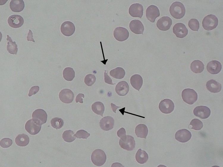
Irregularities
DNA fragmentation can occur under certain conditions in a few different cell types. This can lead to problems for a cell, or it may lead to a cell receiving a signal to undergo apoptosis. Below are a couple of examples of irregular fragmentation that can occur in cells.
1. Red blood cell fragmentation2. Sperm cell DNA fragmentation