
 | ||
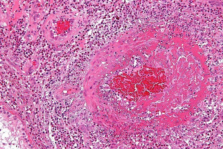
Fibrinoid necrosis is a form of necrosis, or tissue death, in which there is accumulation of amorphous, basic, proteinaceous material in the tissue matrix with a staining pattern reminiscent of fibrin. It is associated with conditions such as immune vasculitis (e.g. polyarteritis nodosa), malignant hypertension, preeclampsia, or hyperacute transplant rejection.
In small vessel vasculitis, fibrin plugs frequently occur in the vessel lumen, but the term fibrinoid is usually used to refer to material outside the lumen of a vessel. Fibrinoid necrosis also occurs in the walls of arterioles in malignant hypertension (blood pressure greater than 200/130 mmHg).
Fibrinoid necrosis is a special form of necrosis usually seen in immune reactions involving blood vessels, known as Type III hypersensitivity reactions. This pattern of necrosis typically occurs when complexes of antigens and antibodies are deposited in the walls of arteries. Deposits of these immune complexes, together with fibrin that has leaked out of vessels, result in a bright pink and amorphous appearance in H&E stains, called “fibrinoid” (fibrin-like) by pathologists. Ultimately, in the living patient most necrotic cells and their contents disappear by phagocytosis of the debris and enzymatic digestion by leukocytes. If necrotic cells and cellular debris are not promptly destroyed and reabsorbed, they tend to attract calcium salts and other minerals and to become calcified.
Common sites undergoing fibrinoid necrosis are, SLE, Aschoff bodies seen in rheumatic heart disease, Arthus reaction, serum sickness, polyarteritis nodosa, poststreptococcal glomerulonephritis, hyperacutely rejected grafts, malignant hypertension (>200/130 mm Hg).