
 | ||
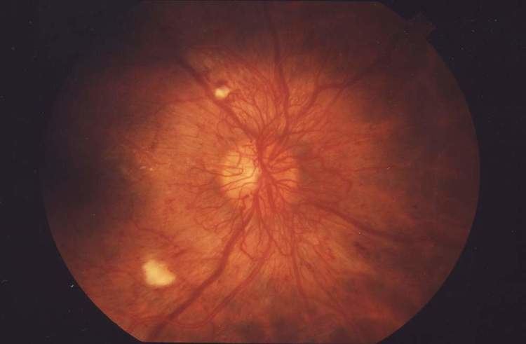
Cotton wool spots are an abnormal finding on funduscopic exam of the retina of the eye. They appear as fluffy white patches on the retina. They are caused by damage to nerve fibers and are a result of accumulations of axoplasmic material within the nerve fiber layer. There is reduced axonal transport (and hence backlog and accumulation of intracellular products) within the nerves because of the ischemia. This then causes the nerve fibers to be damaged by swelling in the surface layer of the retina. A 1981 analysis concluded that "in most instances, cotton-wool spots do not represent the whole area of ischaemic inner retina but merely reflect the obstruction of axoplasmic flow in axons crossing into much larger ischaemic areas". Associated findings include microvascular infarcts and hemorrhages. The appearance of cotton wool spots may decrease over time. Abundant cotton wool spots are seen in Malignant hypertension.
Diabetes and hypertension are the two most common diseases that cause these spots, and the best treatment would be to treat the underlying disease. In diabetes they are one of the hallmarks of pre-proliferative retinopathy. More rarely, HIV and Purtscher's retinopathy can also lead to the appearance of cotton wool spots.
Another condition in which cotton wool spots are found is central retinal vein occlusion.