
Precursor Metanephric blastema MeSH Bowman+Capsule FMA 15626 | Latin capsula glomeruli Dorlands/Elsevier 12211408 | |
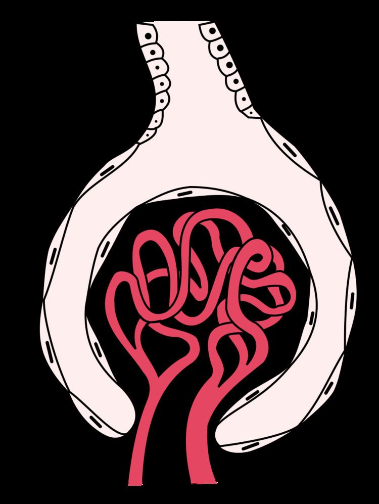 | ||
Bowman's capsule (or the Bowman capsule, capsula glomeruli, or glomerular capsule) is a cup-like sack at the beginning of the tubular component of a nephron in the mammalian kidney that performs the first step in the filtration of blood to form urine. A glomerulus is enclosed in the sac. Fluids from blood in the glomerulus are collected in the Bowman's capsule (i.e., glomerular filtrate) and further processed along the nephron to form urine. This process is known as ultrafiltration. The Bowman's capsule is named after Sir William Bowman, who identified it in 1842.
Contents
Anatomy
Outside the capsule, there are two "poles":
Inside the capsule, the layers are as follows, from outside to inside:
Physiology
The process of filtration of the blood in the Bowman's capsule is ultrafiltration (or glomerular filtration), and the normal rate of filtration is 125 ml/min, equivalent to 80 times the daily blood volume.
Any proteins under roughly 30 kilodaltons can pass freely through the membrane, although there is some extra hindrance for negatively charged molecules due to the negative charge of the basement membrane and the podocytes.
Any small molecules such as water, glucose, salt (NaCl), amino acids, and urea pass freely into Bowman's space, but cells, platelets and large proteins do not.
As a result, the filtrate leaving the Bowman's capsule is very similar to blood plasma (filtrate or glomerular filtrate is composed of blood plasma minus plasma protein i.e. it contains all the components of blood plasma except the proteins) in composition as it passes into the proximal convoluted tubule.
Clinical significance
Measuring the glomerular filtration rate (GFR) is a diagnostic test of kidney function.
A decreased GFR may be a sign of renal failure.
A number of diseases can result in various problems within the glomerulus. Examples include acute proliferative (endocapillary) glomerulonephritis, mesangioproliferative glomerulonephritis, mesangiocapillary (membranoproliferative) glomerulonephritis, acute crescentic glomerulonephritis, focal segmental glomerulonephritis, and diabetic glomerulosclerosis.
Eponym
Bowman's capsule is named after Sir William Bowman (1816-1892), a British surgeon and anatomist. However, thorough microscopical anatomy of kidney including the nephronic capsule was described by the Ukrainian surgeon and anatomist from the Russian Empire, Prof. Alexander Shumliansky (1748-1795), in his 1788 doctoral thesis "De structura renum: Tractatus physiologico-anatomicus" ("About Kidney Structure: an Physiological-Anatomical Treatise," in Latin); thus, much prior to Bowman.
Together with the glomerulus it is known as a renal corpuscle, or a Malpighian corpuscle, named after Marcello Malpighi (1628-1694), an Italian physician and biologist. This name is not used widely anymore, probably to avoid confusion with Malpighian bodies of the spleen.