The body of hyoid bone or central part of hyoid bone is of a quadrilateral form.
Its anterior surface is convex and directed forward and upward.
It is crossed in its upper half by a well-marked transverse ridge with a slight downward convexity, and in many cases a vertical median ridge divides it into two lateral halves.
The portion of the vertical ridge above the transverse line is present in a majority of specimens, but the lower portion is evident only in rare cases.
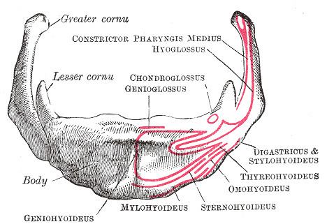
The anterior surface gives insertion to the geniohyoid muscle in the greater part of its extent both above and below the transverse ridge; a portion of the origin of the hyoglossus notches the lateral margin of the geniohyoid attachment.
Below the transverse ridge the mylohyoid, sternohyoid, and omohyoid are inserted.
The posterior surface is smooth, concave, directed backward and downward, and separated from the epiglottis by the hyothyroid membrane and a quantity of loose areolar tissue; a bursa intervenes between it and the hyothyroid membrane.
The superior border is rounded, and gives attachment to the hyothyroid membrane and some aponeurotic fibers of the genioglossus.
The inferior border affords insertion medially to the sternohyoid and laterally to the omohyoid and occasionally a portion of the thyrohyoid. It also gives attachment to the Levator glandulae thyreoideae, when this muscle is present.
In early life the lateral borders are connected to the greater cornua by synchondroses; after middle life usually by bony union.