
 | ||
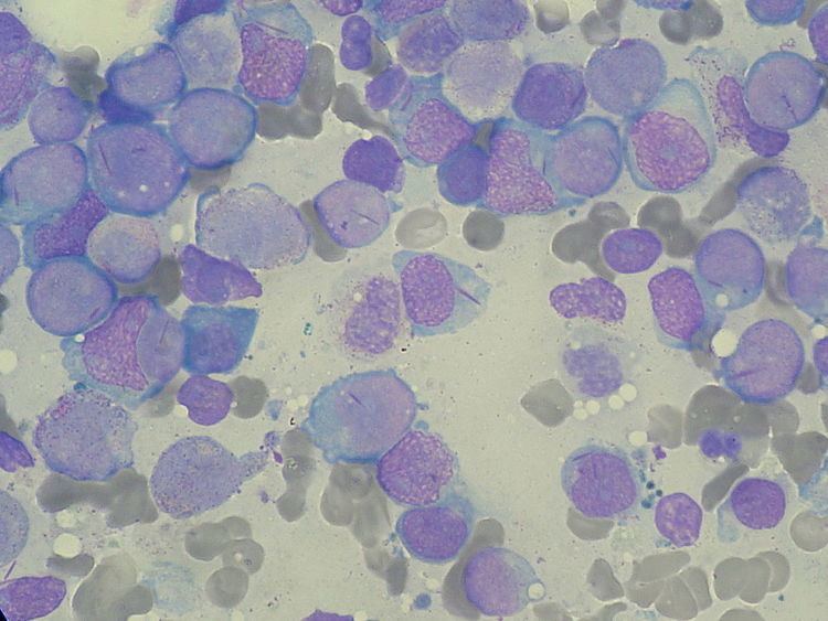
Auer rods are clumps of azurophilic granular material that form elongated needles seen in the cytoplasm of myeloid leukemic blasts. They can be seen in the leukemic blasts of acute myeloid leukemia with maturation and acute promyelocytic leukemia (respectively known as acute myeloid leukemia M2 and M3 in the FAB classification) and in high grade myelodysplastic syndromes and myeloproliferative syndromes. They are composed of fused lysosomes/primary neutrophilic granules and contain peroxidase, lysosomal enzymes, and large crystalline inclusions. Morphologically, the Auer "rods" come in all sizes and shapes. They have been described as needle-shapes with pointed ends (most common), comma-shapes, and diamond-shapes; others were long and rectangular. Occasional corkscrew forms and rare granular Auer bodies were also noted. More appropriately, they can be referred to as Auer bodies.
They are also used to distinguish the pre-leukemia myelodysplastic syndromes: refractory anemia with excess blasts 2 (which has Auer rods) from RAEB 1 (which does not). However, rare cases of RAEB1 show rare Auer rods, and when they do, they have a worse prognosis.
Eponym
These cytoplasmic inclusions are named for John Auer, a US physiologist (1875-1948).
However, they were first described in 1905 by a Canadian physician, Thomas McCrae, then at The Johns Hopkins Hospital, one year before Auer, as acknowledged in Auer's article. Both McCrae and Auer mistakenly thought that the cells containing the rods were lymphoblasts.