
 | ||
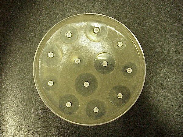
Antibiotic sensitivity or antibiotic susceptibility is the susceptibility of bacteria to antibiotics. Because susceptibility can vary even within a species (with some strains being more resistant than others), antibiotic susceptibility testing (AST) is usually carried out to determine which antibiotic will be most successful in treating a bacterial infection in vivo. Testing for antibiotic sensitivity is often done by the Kirby-Bauer method. Small wafers containing antibiotics are placed onto a plate upon which bacteria are growing. If the bacteria are sensitive to the antibiotic, a clear ring, or zone of inhibition, is seen around the wafer indicating poor growth. Other methods to test antimicrobial susceptibility include the Stokes method, Etest (also based on antibiotic diffusion), Agar and Broth dilution methods for minimum inhibitory concentration (MIC) determination. The results of the test are reported on the antibiogram.
Ideal antibiotic therapy is based on determination of the aetiological agent and its relevant antibiotic sensitivity. Empiric treatment is often started before laboratory microbiological reports are available when treatment should not be delayed due to the seriousness of the disease. The effectiveness of individual antibiotics varies with the location of the infection, the ability of the antibiotic to reach the site of infection, and the ability of the bacteria to resist or inactivate the antibiotic. Some antibiotics actually kill the bacteria (bactericidal), whereas others merely prevent the bacteria from multiplying (bacteriostatic) so that the host's immune system can overcome them. Müeller-Hinton agar is most frequently used in this antibiotic susceptibility test.
An antibiogram is the result of an antibiotic sensitivity test. It is by definition an in vitro sensitivity, but the correlation of in vitro to in vivo sensitivity is often high enough for the test to be clinically useful.
In clinical practice, antibiotics are most frequently prescribed on the basis of general guidelines and knowledge about sensitivity: e.g. uncomplicated urinary tract infections can be treated with a first generation quinolone, etc. This is because Escherichia coli is the most likely causative pathogen, and it is known to be sensitive to quinolone treatment.
However, many bacteria are known to be resistant to several classes of antibiotics, and treatment is not so straightforward. This is especially the case in vulnerable patients, such as patients in the intensive care unit. When these patients develop a hospital-acquired pneumonia, more hardy bacteria like Pseudomonas aeruginosa are potentially involved. Treatment is then generally started on the basis of surveillance data about the local pathogens probably involved. This first treatment, based on statistical information about former patients, and aimed at a large group of potentially involved microbes, is called empirical treatment.
Before starting this treatment, the physician will collect a sample from a suspected contaminated compartment: a blood sample when bacteria possibly have invaded the bloodstream, a sputum sample in the case of a ventilator associated pneumonia, and a urine sample in the case of a urinary tract infection. These samples are transferred to the microbiology lab, which looks at the sample under the microscope, and tries to culture the bacteria. This can help in the diagnosis.
Once a culture is established, there are two possible ways to get an antibiogram:
Once the MIC is calculated, it can be compared to known values for a given bacterium and antibiotic: e.g. a MIC > 0,06 µg/ml may be interpreted as a penicillin-resistant Streptococcus pneumoniae. Such information may be useful to the clinician, who can change the empirical treatment, to a more custom-tailored treatment that is directed only at the causative bacterium.
Often clinical specimens are sent to the clinical laboratory for culture and sensitivity (C&S), which is culture and antibiotic sensitivity testing offered as one combined service. This is because the clinician needs to know (1) which organism is it? and (2) which drugs will work on this strain?